Biopsy and Cyst removal
Biopsy and removal of simple cysts and other lesions of the mouth – Some lesions occurring in the mouth and maxillofacial region required to be examined for any disease. A tissue sample is taken from the lesion and sent for histopathological examination. This procedure is called a biopsy. Some simple lesions and infections including cysts have to be removed.
CASE I
-
-showing-extensive-cyst-in-lower-jaw-middle-region-and-upper-left-jaw.jpg)
3D Cone Beam CT image (CBCT) showing extensive cyst in lower jaw middle region and upper left jaw
-
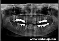
Digital X-ray showing cyst lesions
-
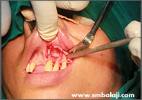
Bony lesion surgically exposed in the upper jaw
-
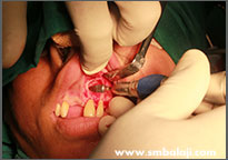
Cyst lesion surgically removed in upper jaw
-
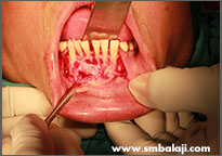
Tori surgically excised from the lower right jaw region
-
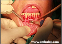
Cyst lesion surgically removed in lower jaw
-
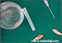
Excised lesions and extracted infected teeth
-
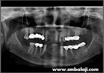
X-ray following surgery showing good bone healing
CASE II
-
-image-showing-impacted-tooth-and-cyst-in-lower-jaw-right-side.jpg)
3D Cone Beam CT (CBCT) image showing impacted tooth and cyst in lower jaw right side
-
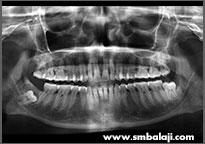
X-ray showing right lower impacted tooth with cyst lesion and other multiple impacted teeth
-
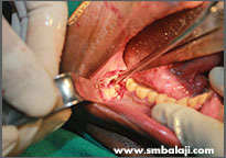
Surgical exposure of impacted tooth
-
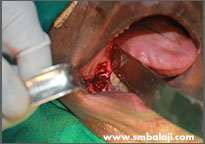
Impacted tooth surgically removed
-
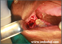
Cyst lesion excised
-
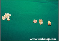
Removed impacted teeth
-
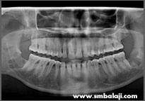
X-ray showing good bone healing after surgery
CASE III
-
-image-showing-impacted-lower-right-wisdom-tooth.jpg)
3D Cone Beam CT (CBCT) image showing impacted lower right wisdom tooth, affected second molar and large cyst lesion in right lower jaw
-
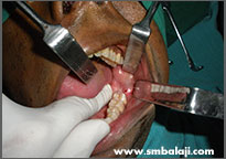
Clinical exposure of the diseased portion of the right lower jaw
-
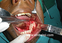
Surgical exposure of the impacted tooth and cyst
-
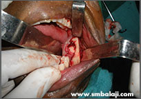
Impacted teeth and cyst excised surgically
-
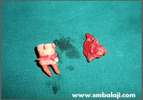
Removed infected tooth and cyst
-
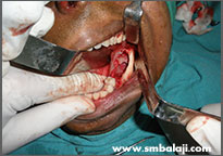
After cyst excision and surgical removal of affected teeth
-
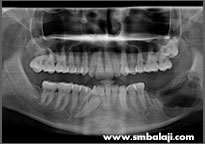
Digital X-ray taken immediately after surgery
-
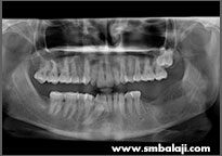
X-ray showing very good bone healing just three months following surgery
CASE IV
-
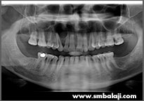
X-ray showing cyst lesion in upper jaw right side
-
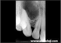
Radiovisuograph showing cyst lesion
-
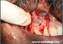
Surgical exposure of cyst
-
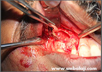
Cyst lesion surgically excised
-
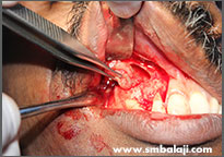
Cyst lesion surgically excised
-
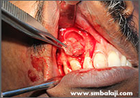
Cyst lesion surgically excised
-
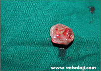
The excised cyst pathology
-
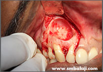
Bone defect cleaned after cyst removal
