Injuries to the jaws and TMJ
Fractures occur in the lower jaw mainly due to road traffic accidents, sport injuries, fist fights and negligence during extraction. The fractures of the lower jaw are classified on basis of the region involved. The diagnosis can be easily made by means of X-rays .The Mandible (lower jaw) is the strongest bone in the face. The tongue and numerous muscles have attachment in the lower jaw.
Upper jaw fracture
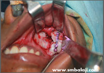
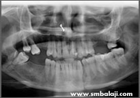
The patient suffered a fracture of his upper jaw bone (Maxilla). As in such cases, the mouth opening gets severely restricted. The fractured site was surgically exposed, the bone fragments were aligned and fixed with bone plate.
-
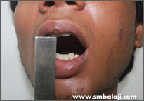
Inability to open mouth due to fractured upper jaw
-
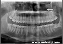
X-ray showing fracture
-
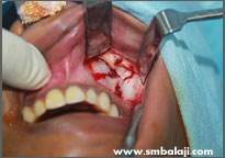
During surgery
-
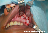
Open reduction of fracture
-
X-ray showing fracture
-
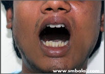
Improved mouth opening postoperatively
Lower jaw fracture
In cases of fractures of the mandible (lower jaw), the fractured segments of bone are stabilized and fixed with bone plates. Subsequent healing of the injury occurs.
CASE I
These are photographs and X-rays of a 45 year old lady who suffered fracture of her lower jaw in 2 regions as a result of a motor vehicular accident. Once identified on X-ray, the fracture site was accessed, and broken fragments of bone were stabilized using bone plates. Following surgery, X-rays taken revealed good healing and recovery.
-
X-ray showing multiple fractures in the lower jaw bone
-
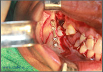
Fractured bone fixed with plate
-
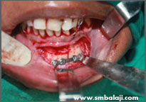
Fractured bone fixed with plate
-
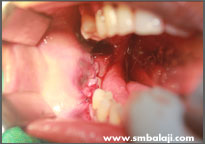
Immediately after surgery
Temporomandibular Joint fracture
Condyle fracture: The condyle is a portion of the lower jaw that forms a part of the temporomandibular joint (TMJ or jaw joint). Depending on the type of impact, the condyle may be fractured either on one side or both. The fractured fragment may simply lie in the jaw joint, just a fracture line can be seen without displacement of the broken bone. Or the broken segments may get disrupted. Treatment varies depending upon the extent of injury and resulting position of the fractured bones.
Dr. S.M Balaji, an expert at treating jaw fractures, emphasizes on a concise algorithm for diagnosis and treatment of these injuries.
CASE 1
In this instance, the patient suffered a condyle fracture. A 3D cone beam CT scan revealed that position of the bone fragment was not overtly disrupted. Such situations warrant a minimal treatment.
After arranging the teeth of the upper and lower law in correct alignment with respect to each other, the occlusion or the way the patient bites is accurately stabilized. To keep the jaws from moving until bone healing occurs, the upper and lower teeth are wired shut. This is called maxillomandibular fixation.
During this time the patient’s diet is restricted to liquid foods. When the jaws are kept stable and immobile, the condyle fracture heals well. After a few days a stable occlusion is achieved and the wiring is removed.
CASE 2
In some cases of trauma, both the right and left condyle may be fractured and displaced. In such instances, the patient’s occlusion or contact between upper and lower teeth gets deranged. The fracture site is surgically exposed, the fractured segments are stabilized and fixed with bone plates. This is done only after accurately restoring the patient’s occlusion.
