Facial symmetry is always considered one of the most influential characteristics for a beautiful face. Although we commonly believe the face to be symmetrical, in reality, typical symmetry is a myth. A noticeably asymmetrical face is considered unattractive or unpleasant and demands cosmetic surgical correction.
Additionally, irregular jaw growth or a crooked face may also cause irregularities in teeth alignment. Corrective and cosmetic jaw surgery techniques give a more symmetrical, aesthetically pleasing face as well as correct functioning. Facial asymmetry is a developmental problem i.e. it occurs due to disturbances in growth and usually manifests in early youth. Facial asymmetry is highly attributed by the underlying facial skeleton and to a larger extent by the morphology of the lower jaw (mandible). Commonly, face asymmetry is because the lower jaw is shorter on one side as compared to the other.
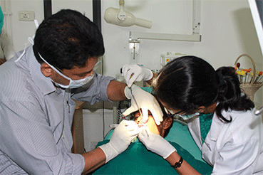
The treatment of facial asymmetry has advanced rapidly over the past two decades. Keeping pace with recent global trends, Balaji Dental & Craniofacial Hospital offers latest advances and sophisticated technologies to attain perfect results. Dr. S.M. Balaji’s expertise is at the forefront in the field of facial asymmetry surgery.
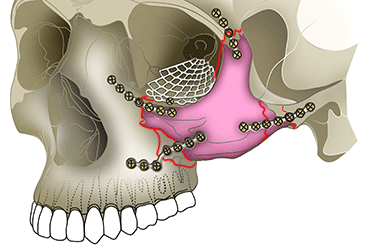
Distraction Osteogenesis is an innovative treatment option in maxilla-facial surgery. In this procedure, the jaw bone is cut on the side it requires to be lengthened and a distractor device is placed. The two arms of the device are attached to the cut bone segments. After a few days the distraction procedure is started. The screw attached to the device is turned gradually (about 1 mm per day) which pushes the bones apart. New bone is formed in the resultant gap. After the desired jaw bone length is achieved, distraction is stopped. Once the new bone stabilizes, the distractor device is removed.
Distraction is the magical technique by which we can achieve bone growth even in adulthood. The versatility of this technique lies in the fact that corrections can be achieved more precisely even to the last millimeter.
Besides unequal jaw growth, there are other rare causes of facial asymmetry. Inspite of normal articulation of jaws, soft tissue discrepancies such as masseter hypertrophy (enlargement of masseter – a muscle that facilitates chewing) and muscle deficit on one side of the face (as seen in some syndromes like Parry-Romberg syndrome) also result in facial asymmetry.
In rare instances, the masseter is enlarged either on one or both sides. Most commonly, patients complain of facial asymmetry. Comprehensive treatment includes surgical reduction of the masseter muscle to make the face more symmetrical and enhance the esthetics of the face.
In patients suffering from rare conditions like Parry-Romberg syndrome, there is degeneration and shrinkage of the soft tissues generally on one side of the face. This leads to facial asymmetry. To treat this, the patient’s fat tissue is grafted and placed beneath the skin on the deficient side of the face. Following surgery, the patient’s face appears more symmetrical and balanced.
Facial asymmetry in a patient with Hemifacial Microsomia
Ear deformity in patient with hemifacial microsomia
Jaw bone segmented on the left side and preparation to fix distractor device
Distractor device fixed to jaw bone
Ear deformity in patient with hemifacial microsomia
Jaw bone segmented on the left side and preparation to fix distractor device
Activating the distractor to lengthen the jaw bones
Bone segments moving apart to lengthen bone
Facial asymmetry corrected with distraction osteogenesis
This reconstruction is a surgical procedure in which a graft consisting of a section of the skin along with blood vessels is lifted from the forehead region with the base of the graft still attached to the forehead in between the eyebrows. The wound on the forehead is sutured. The graft is then brought downwards and attached to the nose tip.
After blood supply is established in the nose, the graft tissue is cut off from the forehead. With this reconstruction, there is no color mismatch and it gives the nose a more normal appearance. Along with this if required, cartilage grafts from the ribs can be taken to reconstruct the side or central prominence of the nose. The type of reconstructive surgery required will depend on the extent of deformity of the nose.
A social worker and human rights activist was referred to Balaji Dental and Craniofacial Hospital for specialized reconstructive surgery of the nose. His work involved him to interact with many people from different walks of life. In one such encounter, during a civil unrest, a miscreant bit his nose. After initial first aid treatment, he was referred here for expert care.
The photographs of a 7-year-old boy show how the nose injured in an accident was reconstructed using a graft from the forehead. The sharpness of the nose and nose tip is accurately reconstructed to give a more natural form and appearance. The photographs of a 20-year-old girl show how the shape of the nostrils was deformed since birth. The side of the nose is rebuilt giving a more normal structure and appearance.
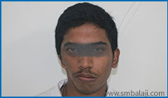
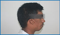
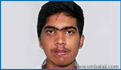
A 22 year old male reported to our hospital for the surgical correction of asymmetry over the left side of the face. Facial asymmetry may be present in cases of hemifacial microsomia, TMJ ankylosis or may have resulted following condylar fractures. This patient had a mandibular deficiency at the ramus level due to condylar fracture and deficiency measured almost 17 mm with a resultant occlusal cant.
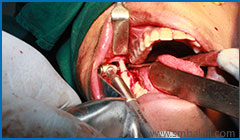
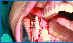
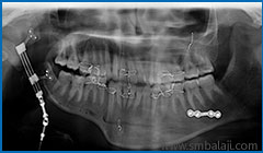
After thorough clinical and radiological examination Maxillofacial Surgeon Dr. S. M. Balaji planned to correct the facial asymmetry using internal distraction osteogenesis with maxillomandibular distractor. Paragingival incision was placed over the left angle region along the anterior border of ramus. Full thickness mucoperiostal flap was reflected buccally and bone exposed. The direction of the osteotomy cut and positioning of the distractor is the most important as it determines the vector of distraction and this determines the direction of the bone growth. The distractor device was positioned and fixed using screws. Osteotomy is then completed along the medial cortex. Distractor device was checked and wound closure done. Le Fort I osteotomy was completed in maxilla. After a latency period of 5 days, distraction at the rate of 1 mm per day was accomplished and facial asymmetry was successfully corrected.

Preoperative facial view showing aymmetrical appearance of the face due to reduced height on the left side
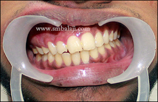
Preoperative bite showing occlusal cant due to reduced mandible height on the left side
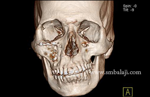
Preoperative 3D CT scan showing altered height of the ramus of the mandible on the left side leading to asymmetry
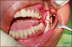
During surgical procedure, distractor device fixed to the outer surface of the ramus with correct orientation with the jaw
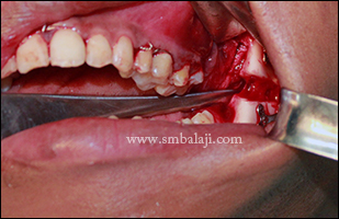
Trial activation done to check the movement of the mandible
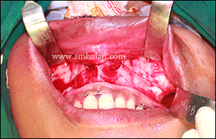
Lefort I osteotomy cut was placed in the maxilla and fixed to the mandible by IMF for simultaneous distraction
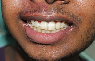
Postoperative view showing corrected occlusal cant by lengthening the lower half of the face using distraction osteohistogenesis