Hemifacial Microsomia is a birth deformity affecting the bony skeleton and soft tissues of the craniofacial region. It is typically characterized by pronounced asymmetry of the face due to deficient growth of the lower jaw (mandible) on one side, deficient soft tissues of the face and underdeveloped ear (microtia).
Bony and soft tissue structures on one side of the face grow lesser than the other side. The affected side of the face appears disproportionately smaller than the other. Commonly affected are the lower jaw, eye, ear, facial nerve and cheek. The most significant of these deformities is that of the jaws, which when corrected, minimizes the entire deformity and makes the face look more symmetric.
In hemifacial microsomia, the shape and size of the lower jaw is deformed on one side causing an asymmetric face. The chin appears deviated to the affected side. Facial asymmetry progresses as the patient grows. As a result, teeth bite (occlusion) is slanted.
Based on various parameters like severity of the defect and age of the patient, there are different treatment options. Cheek and eye socket bone defects if present, can be corrected with specialized craniofacial surgeries. Ear and soft tissue defects can be rehabilitated with reconstructive surgeries.
The goal of treatment in hemifacial microsomia is to elongate the deficient jaw bone to restore facial symmetry and correct the slanting bite (occlusion). To achieve this, an advanced and effective treatment technique is distraction osteogenesis. This is a new technique for regenerating new bone by slow, progressive stretching of the bone, without requiring a bone graft.
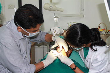
The jaw bone on the deficient side is cut. A sophisticated device called distractor is placed such that the two arms of the device are fixed to the two segments of jaw bone. After a few days, a screw attached to the distractor is turned gradually, ideally at a rate of 1 mm per day. When this is done, the two cut segments move apart and new bone is formed in the resultant gap. After the new bone is stabilized, the distractor device is removed.
Subsequently, the jaw bone is lengthened to the desired amount correcting the asymmetry of the face. This is a powerful tissue engineering technique for generating unlimited bone.
Eminent Facio-Maxillary Surgeon Dr. S.M. Balaji is highly acclaimed for his prowess in the treatment of Hemifacial Microsomia. He is a pioneering expert in this field and has applied several revolutionary techniques including “Simultaneous Maxillary and Mandibular Distraction” giving excellent results to set right facial asymmetry.
Our hospital has to its credit the maximum number of cases of Hemifacial Microsomia successfully treated with Distraction Osteogenesis (Distraction of both upper and lower jaw) for the correction of facial asymmetries.
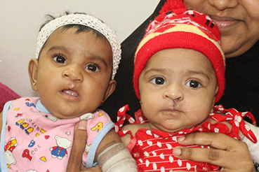
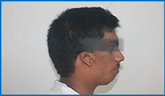
Facial asymmetry in a patient with Hemifacial Microsomia
Ear deformity in patient with hemifacial microsomia
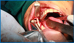
Jaw bone segmented on the left side and preparation to fix distractor device
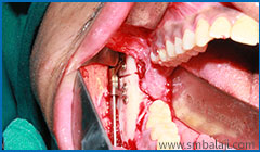
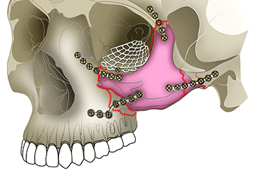
Distractor device fixed to jaw bone
Ear deformity in patient with hemifacial microsomia
Jaw bone segmented on the left side and preparation to fix distractor device
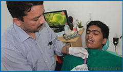
Activating the distractor to lengthen the jaw bones
Bone segments moving apart to lengthen bone
Facial asymmetry corrected with distraction osteogenesis

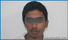
Hemifacial Microsomia with facial asymmetry, slanting smile and chin deviation

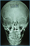
CT scan image showing asymmetry of facial skeleton

Improper bile due to hemifacial microsomia

Distraction Osteogenesis treatment planning

Placing the intraoral distractor

Fixing the intraoral distractor during surgery

Bite improved after treatment
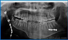
X-ray showing skeletal asymmetry of the face corrected after treatment

Asymmetry of the face corrected with distraction osteogenesis without scars/p>

Asymmetrical face in a patient of hemifacial microsomia

CT scan image before surgery showing facial asymmetry

X-ray showing deficient growth of the lower jaw bone on left side

X-ray at the end of distraction treatment showing corrected asymmetry

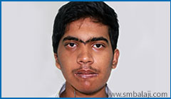
Face made more symmetrical after distraction osteogenesis