Life-saving jaw surgery for Chinese baby from Singapore suffering from potentially fatal facial bone condition & Respiratory distress- using advanced Distraction Osteogenesis technique
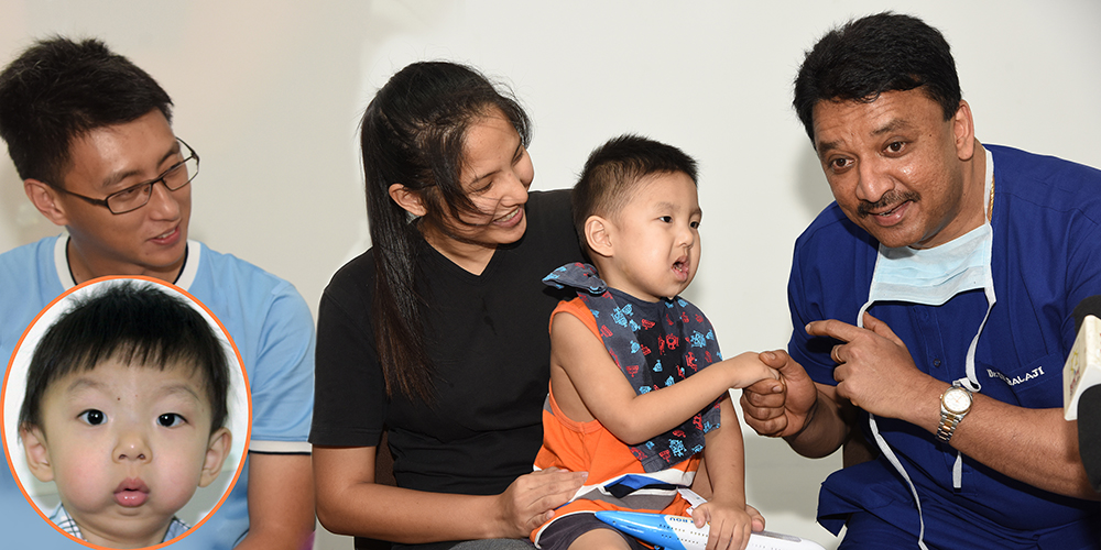
2-year-old boy Chayce Lee was brought to our hospital by his father Mr. Lee Teck Chuan and mother Mrs. Tan Vie Vian, both employed as Sales Executives in Singapore, for saving their son’s life. The parents had sleepless nights and were very scared because their baby had severe breathing difficulties. They approached many doctors in Singapore, Kuala Lumpur, Hong Kong and other places but the doctors gave different opinions and the parents were not satisfied. The doctor refused to operate because of the baby’s challenging condition. Finally the parents approached us and discussed with Dr. S. M. Balaji about their baby’s life-threatening condition, they were convinced that this is the best center to get their son treated.
Case History
The baby was born with rare autosomal recessive genetic disorder called Pierre Robin syndrome, a potentially life-threatening condition with severe abnormalities affecting the head and face. This is a very rare condition which affects only about 1 in 10000 live births.
Pierre Robin syndrome: This is characterized by very small lower jaw (micrognathia) that is very backwardly placed (retrognathic), receding chin and tongue that is placed much further back than normal (glossoptosis). This is a very serious condition because the lower jaw is severely underdeveloped, there is very less tongue-space, the tongue keeps falling-back in the throat blocking the airway completely. The combination of these features can lead to difficulty in breathing and problems in eating. Because of respiratory distress, low blood oxygen and brain damage (due to difficulty breathing) may occur. As a result, some affected babies have an inability to grow and gain weight at the expected rate (failure to thrive). Earlier this condition was treated by stitching the tip of tongue to lower lip to prevent tongue fall-back into the wind-pipe but this had many disadvantages.
The baby had Obstructive Sleep Apnea (breathing & sleeping distress) wherein breathing repeatedly stops for a few seconds to minutes. The child used to wake up often gasping for breath. His breathing used to stop repeatedly and start and the pauses used to last from a few seconds to minutes. This could occur 30 times or more an hour. Typically, normal breathing then starts again with a loud snort or choking sound. The parents were very worried for their baby’s life. Also the boy had heavy snoring, difficulty in swallowing (dysphagia) and problems in speech.
Added the boy had a severe fall injury few months after birth which caused his jaw joints (temporomandibular joints TMJ) to became fused to the skull bone on both sides (bilateral TMJ ankylosis). Since the potential growth center of the lower jaw is the TMJ, this problem should also be addressed later. Moreover by birth the boy has an orthopedic condition of the hip called Tom Smith arthritis.
Balaji Dental and Craniofacial Hospital
Dr. Balaji assessed his face bone deformity using advanced 3DCT scan. To rescue him from this life-threatening condition and to improve his breathing, Dr. Balaji performed mandibular advancement using Pediatric Distraction Osteogenesis to bring the lower jaw forward in a slow, controlled manner, avoiding bone grafting from anywhere in the body, inside the mouth itself (intraorally) without any scars.
The child’s condition required lengthening of the lower jaw bone. One of the ways to do this is by taking a bone graft from the rib or hip. But in this case owing to the very young age of the boy, graft could not be taken.
To stabilize the breathing Tracheostomy was done in which a small hole was made in the wind pipe (trachea). Using advanced Pediatric Mandibular Intraoral Distraction Osteogenesis, the lower jaw bone was cut at the angle on both sides and the distraction devices fixed to the bone. After a latency period of 5 days, the device was gradually activated which caused the bone to expand in 3 dimensions. At the rate of 1 turn, half mm of new bone was grown automatically over 50 days achieving 2.5- 3.00 cm of bone growth and ultimately the lower jaw was brought forward. The uniqueness of the correction is that the distractors were placed inside the mouth (intraorally) so there are absolutely no scars on the face and existing native bone growth was induced.
Successfully as the new jaw bone grew automatically every day, the lower jaw was brought forward, fall back of the tongue & snoring problem gradually stopped and the boy is now able to breathe and swallow easily. Moreover the entire procedure was done inside the mouth so there is no scar on the face. His parents are very happy and relieved. Following this surgical procedure to gain normal breathing, the boy is now out of emergency condition and he can breathe easily without snoring. After consolidation period of 3 months, the surgery to remove the distractor devices was done and the parents are very happy and will be returning to their homeland. They were informed that at the age of 7 years, the TMJ ankylosis will be surgically corrected with Growth Center Transplantation (Costochondral junction being junction of the bone and cartilage from the rib) which will automatically induce normal growth of the lower jaw.
Recently, an 18-month-old Pakistani toddler from Karachi born with a congenital, bilateral Tessier’s facial Cleft Type 4 with orbital (eye) bone clefts was successfully rehabilitated at Balaji Dental and Craniofacial Hospital.
Non-fusion of facial bones including the bones forming the eye sockets (orbit) results in facial clefts. Of the nearly 20 major types of facial clefts, those involving orbit are often rare (more than 1 in 100,000 live births) and rehabilitations are complex.
Children born with such severe deformities suffer immense difficulties in feeding, breathing, speaking and psychological trauma because of their appearance. Master Noman Arshad was born to Pakistani farmhands, Mr. Arshad Ali and Mrs. Raziya. Their 18 month old son, Master Noman was born with this bilateral Tessier’s facial Cleft Type 4 defect. Mr. Arshad Ali sought the best treatment for his son's defect. His search led him to renowned Dental Surgeon Dr. Asif Arian who is the President of Asia Pacific Dental Federation (APDF) & Former President of Pakistan Dental Association. Being aware of Chennai based Craniofacial Surgeon Dr. S. M. Balaji’s work, Master. Noman was referred to Balaji Dental and Craniofacial Hospital.
It was very tough to bring the Pakistani boy to India. Dr. Balaji took rehabilitation as a challenge at several levels. The defect in itself was complex and it required skill and precision as the challenge was to salvage some vision for the boy. Dr. Balaji evaluated the extent of facial cleft with the help of 3D CT scan. The boy had bilateral cleft lip & palate which was extending to the orbital (eye) floor resulting in a deformed eye and eyelids. The left eye was congenitally absent. There was no vision in the left eye and also the right eye had only 40% vision. Due to the extensive involvement of the eyes in the cleft defect, the nasolacrimal ducts were damaged causing epiphora or continuous tear flow from the eyes. Because of the cleft, the child could not be normally fed nor was there a chance of good speech formation. The central part of cleft was attached to the upper lip.
Dr. Balaji’s plan was to immediately close the lip defect to recreate the mouth to enable the baby to be fed well and to have a reasonable facial profile. In addition, Dr. Balaji’s foremost concern was to save the baby’s vision in the right eye, prosthetically replace the congenitally missing left eye to improve the appearance for social acceptance and to correct the abnormal tear flow. Hence in a 6 hour long surgery, Dr. Balaji closed the bilateral cleft lip. He then recreated the upper eyelids. All extra tissue tags were carefully dissected out. The internal architecture of the eye socket was recreated. In the process, careful surgical handling ensured salvaging of the vision of the right eye. A prosthetic eyeball was fixed in the left eye to closely resemble a normal eye. The abnormal tear flow was corrected with DCR or Dacryocystorhinostomy in which a new direct communication was created by placing a silicone tube to enable the tears to drain into the nose.
With the completion of first step of reconstruction itself, Master Noman now has a more normal appearance and his feeding has improved greatly. The parents are now psychologically strengthened with their baby’s improved facial appearance & health and they are returning to Karachi. This being a unique case of a Pakistani baby successfully receiving facial reconstruction surgery in Chennai, a press meet has been organized. The awareness of availability of corrective methods for such rare facial deformities in Chennai needs to be created.

